Showing posts with label ORTHO PHYSIO. Show all posts

What is ORTHO CLINCAL EXAMINATION Hip Joint and treatment management physiotherapy


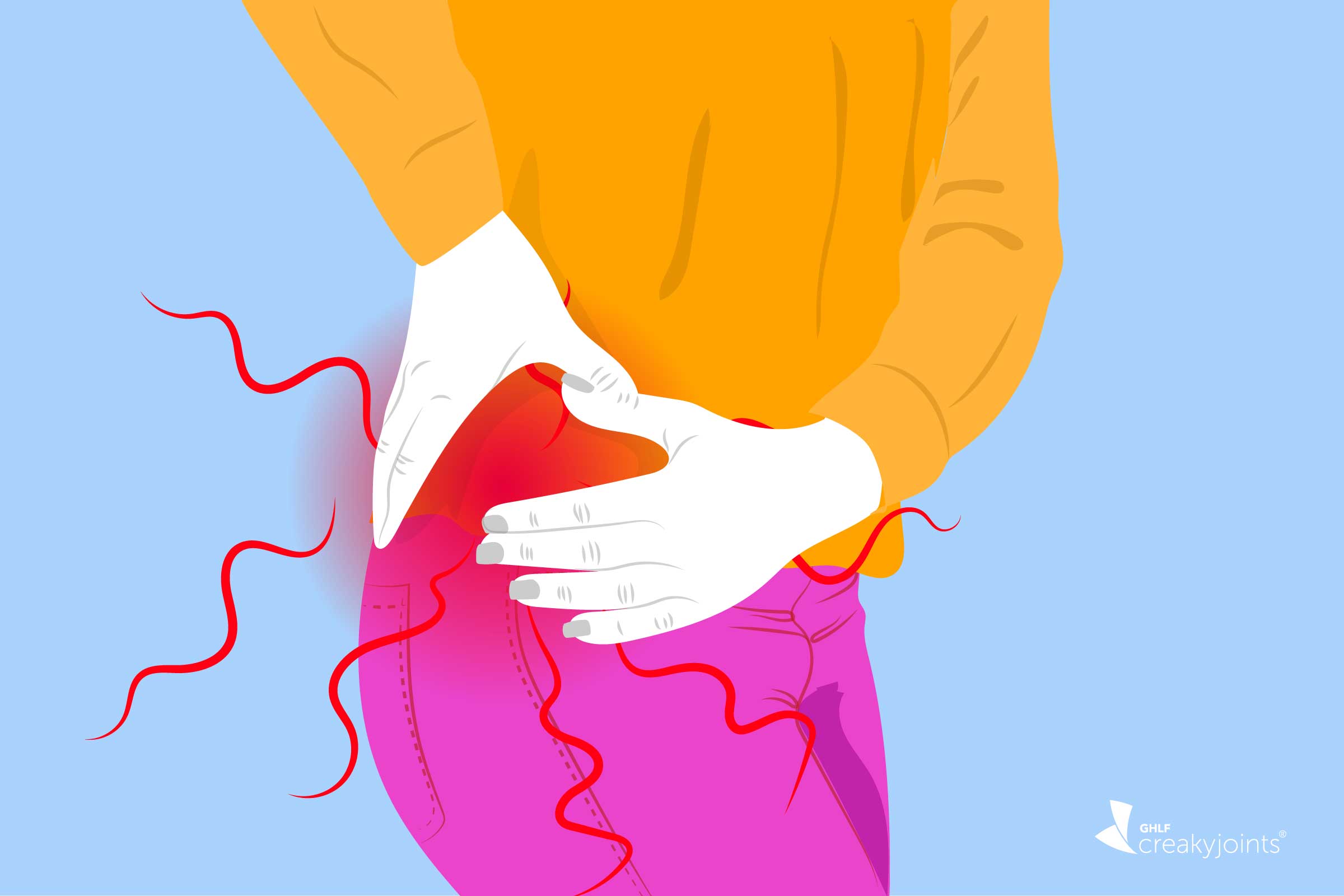
Arthritis in the Hips: Symptoms, Types of Hip Arthritis, Treatment

Introduction


Briefy explain to the patient what the examination involves
Ask the patient to remove their bottom clothing, exposing the hip
Offer the patient a chaperone, as necessary
Always start with inspection and proceed as below unless instructed otherwise; be prepared to be instructed to move on quickly to certain sections by the examiner.

Inspection
Introduce yourself to the patient
Wash your hands
Whilst the patient is standing:


Assess patient gait, such as
Trendelenburg gait
 
 – caused by dysfunction of the hip abductors (gluteus medius and minimus), the patients contralateral hip drops when walking; the patient often offsets this by leaning their trunk toward the affected hip
Antalgic
 
 – produced from weight bearing on painful leg, resulting in a shortened stance-phase and producing the characteristic ‘limping’ patient
Examine for quadriceps muscle bulk
Ask the patient to lie supine on the bed:

Assess for:
Skin changes (uncommon in primary hip pathology as the joint is deep)
Scars (indicative of previous surgery)
Swelling (also uncommon, as the joint is deep)
Measure leg length with a tape measure. This assesses whether there is an actual leg length discrepancy and whether there is any pelvic tilt present to compensate for this:
True leg length = ASIS to medial malleolus
Apparent leg length = pubic symphysis to medial malleolus 

Palpate
Assess for temperature
Feel for trochanteric bursa tenderness
Palpate over the greater trochanter

Movement
All movements are passive when examining the hip, ensuring to note any pain, the range of motion, and any crepitus.

Abduction and adduction
Place one hand across the patient’s pelvis to ensure that the pelvis remains still and that the movement is coming from the hip joint and not the pelvis
Flexion and extension
Internal and external rotation (assessed with the hip flexed)

Special Tests

Thomas’ Test (assesses for fixed flexion deformity)
 

Have patient lying in the supine position, and place one hand underneath the patients lumbar spine to ensure loss of the lumbar lordosis
Fully flex the contralateral hip and observe the ipsilateral hip (i.e. the one that you are examining). Any flexion in this hip suggests a fixed flexion deformity. Repeat this test on both sides
Trendelenburg test (assesses abductor muscle function)*
 

Ask patient to place their hands on your outstretched hands (for stability) and ask them to stand on the leg that you are examining, lifting the contralateral leg off the ground (for 30 seconds).
Feel for a drop in the pelvis on the contralateral side. If there is abductor pathology (gluteus medius and minimus) on the side you are examining then the contralateral side (the normal side) will sag down (“Sound Side Sags”)

What is ORTHO CLINCAL EXAMINATION OF HAND and management 🙌


Hand model - Wikipedia

Introduction
Introduce yourself to the patient
Wash your hands
Briefly explain to the patient what the examination involves
 

Ask the patients to place their hands on a pillow
Always start with inspection and proceed as below unless instructed otherwise; be prepared to be instructed to move on quickly to certain sections by the examiner.

Inspection
Ask the patient to pronate their hands

Inspect for pathology in the:
Nails
Heberdens (DIPJ) or Bouchards (PIPJ) nodes (present in osteoarthritis)
 

Dorsum of the hand
Check the rotational axis of each finger (look from the tips of the fingers) to assess for any rotational deformity
Ask the patient to supinate their hands

Assess for any obvious pathology in the palms (e.g. Dupuytren’s contractures)
Ask the patient to flex their elbows and show you their elbows / dorsal surface of their forearms

Check for psoriatic plaques, rheumatoid nodules, or gouty tophi
Palpation
Ask the patient to again place their hands back on the pillow, in a supine position:

Assess temperature
Using the dorsal surface of your own hand,
 feel distal to proximal along the patient’s hand Mumbai's maximum temperature rises to 34° C | Mumbai | Mumbai Live
 and forearm, and compare
 with the contralateral side
Feel for the radial pulse
Assess the muscle bulk of the thenar 
eminence and hypothenar eminence
Run the pad of your thumb firmly over these areas
Compare for asymmetry, caused commonly by disuse or denervation
 (i.e. carpal tunnel syndrome)
Assess the tendons of the hand, feeling for nodules or contractures
Ask the patient to pronate their hands:

Palpate the bony anatomy of the hands, feeling for any tenderness
The radial and ulnar styloid processes
The carpal bones
Along the length of each metacarpal
Gently squeeze all 4 MCP joints together (often painful in inflammatory arthropathies)
Bimanually palpate all of the MCP joints, PIP joints, and DIP joints
This is best done by placing the thumb and index finger around the joint in a pincer like grip, and the other hand the same at 90o to the first hand, feeling for tenderness and laxity in the joint
Movement
Ask the patient to keep the hands in the pronated position:


Check the extensors of the hand, asking the patient to
Extend the wrist against resistance (extensors)
Raise the thumb off of the pillow (EPL)
Hold the wrist passively in extension and ask the patient to extend their fingers (ED)
Flex the fingers to 90oat the MCP joints and ask the patient to extend their fingers again (lumbricals)
Ask the patient to supinate the hands:

Check the flexors of the hand, asking the patient to
Flex the wrist against resistance
Flex the fingers at the PIP joints
Isolate the 3 fingers that are not being tested (simply hold them in the natural anatomical position) and ask the patient to flex the finger being tested (FDS)
Flex the fingers at the DIP joints
Isolate the proximal and middle phalanges by holding them firmly and then asking the patient to flex the distal phalynx of that finger (FDP)
Check the action of the wrists, asking the patient
Abduct their wrists against resistance
Adduct their wrists against resistance
Check the action of the thumbs, asking the patient to
Flex the thumb (FPL)
Abduction the thumb (APL and APB)
Adduction the thumb (AP)
Opposition the thumb (OP)
Assess the functioning of the hand by assessing:

Power grip
Place two fingers in to the patient’s palm and ask them to squeeze as firmly as possible
Pincer grip
Ask the patient to pinch the tip of you finger
Fine motor function
Ask the patient to pick up a penny or do up the buttons on a shirt
Check the sensorimotor function of the terminal branches of the brachial plexus*:


Median nerve
Motor – confirm thumb abduction is present (tests APB)
Sensation – check at the radial border of tip of index finger
Radial nerve
Motor – confirm MCPJ extension is present (tests ED)
Sensation – check at the dorsal surface of first digital web space
Ulnar nerve
Motor – confirm finger abduction & adduction (tests palmar and dorsal interossei)
Sensation – check at the ulnar border of tip of little finger
Assess the vascular status of the hand by assessing:

Colour / temperature / capillary refill time
*In the context of trauma, a different assessment is performed, assessing each aspect of the brachial plexus in turn

Special Tests


Phalen’s Test (test for carpal tunnel syndrome)
 

Ask the patient to place the dorsal surfaces of their hands together and then fully flex the wrists (the ‘reverse prayer’). Hold for 30–60 seconds. This will reproduce their symptoms in positive cases
Tinel’s Test (test for carpal tunnel syndrome)
 

Gently percuss over the volar aspect of the carpal tunnel – this is found just distal to the wrist crease, overlying the carpal bones. In a positive test, the patient will report paraesthesia in the distribution of the median nerve
Finkelstein’s Test (test for DeQuervain’s Tenosynovitis)
 

The thumb is flexed across the palm and the fingers are then wrapped around it. The wrist is then passively adducted (or ulnar deviated). This causes a disproportionate amount of pain over the radial styloid on the affected side

Assess again by running down with the pad of your thumb